


Radiology is a branch of medicine that utilizes medical imaging techniques to diagnose and treat diseases and injuries. Radiologists are medical doctors who specialize in interpreting medical images obtained through various imaging modalities. Radiology plays a crucial role in modern medicine, providing valuable information for diagnosing conditions, planning treatments, and monitoring patients’ progress. Here are some key aspects of radiology:
Imaging Department has the following services:
- Digital X-Ray
- Digital Mammography and Tomosynthesis
- Sonography and Color Doppler
- CT Scan
- MRI
- Interventional Radiology
- PET CT
DIGITAL X-Rays
There is one fixed high multifrequency x-ray machine dedicated for Radiography and Fluroscopy. Digital Radiography (DR) Detectors are used to obtain Digital X-Rays. All forms of conventional X-Ray are taken; long length imaging is also performed with Digital stitching. This is useful for scoliosis as well as evaluation of angulation and limb length in patients with marked osteoarthritis of the knees. All fluoroscopy procedures such as barium swallow, meal, follow through, enema, Intravenous Urography, micturition cystography, retrograde urography, hysterosalpingography, sialography, fistulograms are performed.
Digital portable x-rays –
These are performed at the bedside, in operation theaters. There are 2 portable x-ray machines, which use digital Digital Radiography(DR)Detectors. The use of Digital Digital Radiography (DR) Detectors results in pristine image quality as well as considerable reduction in radiation exposure which is a first in the country.
CARM –
There are 2 CARM machines, which are used in the operation theatre, ICU, Endoscopy department. They are useful for imaging during procedures helping guidance of the procedure under image intensifier control thus reducing complications and procedure time.
Radiation Protection –
Synergy Institute of Cancer Care and Research is extremely sensitive to the fact that x-ray machines emit Radiation. This radiation is extremely safe and no conclusive studies have proven harm to humans. Even then we take extreme care. All our x-ray machines are low radiation output machines.
MAMMOGRAPHY
Mammography machines are used to detect breast cancer at an early stage before it becomes palpable. Also mammography is useful in evaluating a palpable lump. Breast cancer is one of the most common cancers in women today. With increasing incidence of breast cancer, mammography is recommended as a part of health checkup for women. It has been established that early detection leads to very high cure rate and excellent prognosis.
SONOGRAPHY
Sonography one of the basic and essential diagnostic modality. These High end ultrasound machines have excellent image quality and are equipped with modern applications like elastography, Harmonic Imaging and 3D imaging. The Sonography machines are equipped with a wide range of transducers so all types of Sonography examination can be performed. Routine, endocavitatory, high resolution, 3D and doppler’s of any vessel are done. Portable Sonography is performed at the bedside for inpatients who cannot be transported to the imaging department. These patients also benefit from the high-resolution machines at the bedside.
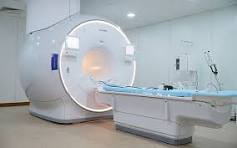
MRI
MRI is the greatest medical advancement of all time. A MRI machine uses a very strong magnetic field – 30,000 times that of the earth. With the use of coils, gradients and an extremely powerful computer it produces amazing high resolution crystal clear images of all regions of the human body. MRI is absolutely safe, there is no radiation nor any adverse effect on the human body. Occasionally patients may feel a bit warm during the san in the region where the scan is being performed. Patients with pacemakers, metallic aortic valves, infusion pumps and cochlear implants should not undergo MRI studies. Some pacemakers are MRI compatible but only to 1.5 Tesla and not 3Tesla. Some metallic aortic valves are also safe. Please carry details of your metallic valve to know if it is safe.
INTERVENTIONAL Radiology
Imaging is very useful in acting as a guide to obtain material for microbiology as well as histopathology. The main advantage of Guided biopsy is that the whole tract of the needle is under constant supervision and the sample material is obtained from the precise part of the lesion while simultaneously avoiding all nearby arteries and organs. This leads to a much safer procedure and a drastic fall in complication rates. The quality of sample material collected is also vastly improved.
The main contraindication relates to bleeding abnormalities. An INR beyond 1.4 and or a platelet count less than 70,000 are considered contraindications. If the biopsy is necessary for further management, platelet transfusion and fresh frozen plasma may be used to minimize the risk of bleeding. Patients on aspirin or antiplatelet agents should withdraw these drugs 3 to 5 days prior to any intervention.
CT SCAN-
This CT scanner also performs all CT scan procedure such as CT scan of head, neck, paranasal sinuses, chest, abdomen, pelvis and bones. As it is a very high end scanner it is very good at performing angiography of any region of the body, head, neck, pulmonary, aorta, renal, upper and lower limb.
PET CT
Positron emission tomography with computed tomography (or PET-CT as it is commonly known as) is an advanced imaging technique used to study various diseases. PET scan is a 3D image obtained from studying functions of human body at the cellular levels. This image is then fused to structural information obtained by a CT scan, giving us the best of both worlds, a fused functional-anatomic image..
Basics of PET-CT hat is a PET-CT scan ?
It is an abbreviation of ‘Positron emission tomography with computed tomography’. It is the imaging of the function and activity of tissues in the body based on an injection of a radioactive medicine. The tracer, most of the times, a radioactive form of glucose, is usually injected intravenously. This tracer goes to most of the cells in the body as a normal distribution, but accumulates more in cells with higher activity, which usually corresponds to the site of disease. On the PET scan these are seen as bright areas.
What are the common indications?
Cancers:
Cancer cells light up bright on the PET scan because they have higher metabolic activity as compared to normal cells, thus enabling to pick disease with higher sensitivity. PET scans are of immense value in
- detecting cancer
- Assessing if the cancer has spread
- In an on-going therapy, checking if the treatment is effective
- To assess response to therapy after completion of the therapy
- Surveillance to see if the disease has recurred
PET scans however must be read with caution since many non-cancerous but active disease cells also can show activity on the scan.
Brain disorders
PET scans can be used in the evaluation of certain brain disorders like
- Dementias like Alzheimer’s disease
- Epilepsy and seizure disorders
- Brain tumours
What are the risks involved ?
For the PET-CT scan, a radioactive medicine will be injected in the body. This does not have any side effects since it is a radioactive form of the glucose. Additionally, the amount of radiation you are exposed to is very minuscule to cause any side effects. However it is not advisable to get the scan done if you are pregnant or in on-going breast feeding. Speak to your doctor if either of these conditions exist.
The CT portion of the PET-CT involves administration of intravenous iodinated contrast, which is the same medicine that is used in a conventional CT scan. There would be no major effects in majority of the cases with the exception of some burning sensation at the site of injection or some nausea. Additional uncommon but documented side effects are vomiting, Rare allergic reactions are also known, may manifest in the form of rashes and chills, in some instances with transient breathing difficulty. These are usually manageable with simple solutions and drugs.
